Rib Cage Muscles Diagram / Ultrasonographic Assessment Of Parasternal Intercostal Muscles During Mechanical Ventilation Annals Of Intensive Care Full Text : The other attachment of these muscles is usually considered to be either superior or inferior to the rib attachment.
byAdmin-
0
Rib Cage Muscles Diagram / Ultrasonographic Assessment Of Parasternal Intercostal Muscles During Mechanical Ventilation Annals Of Intensive Care Full Text : The other attachment of these muscles is usually considered to be either superior or inferior to the rib attachment.. Thoracic cage is a skeletal framework which supports the thorax. The following general rules regarding actions can be. Identify muscles of the rib cage. These rib muscles automatically get worked when you do bench presses, push ups and dips, but a few bonus exercises can help you really zero in for a more chiseled torso. For more anatomy content please follow us and visit our website:
Best viewed on 1280 x 768 px resolution in any modern browser. It is formed by the vertebral column, ribs, and sternum and encloses the heart and lungs. The ribs are a set of twelve paired bones which form the protective 'cage' of the thorax. It provides a strong framework onto which the muscles of the cramps in ribcage are often observed in those who strain or overwork their upper body. At the chest many rib bones connect to the sternum via costal cartilage segments of hyaline cartilage that allow the rib cage to expand during respiration.
8 Muscles Of The Spine And Rib Cage Musculoskeletal Key from musculoskeletalkey.com When you exhale, the rib cage moves down again, squeezing the air. The muscles of the thoracic cage are the pectoralis major, pectoralis minor, serratus anterior, subclavius, intercostal (external, internal and innermost) the subcostal muscles are strips of muscle located on the internal surface of the lower ribs, sharing a plane with the innermost intercostals. The function of the rib cage is to filter the blood it receives, processing the blood. 16 photos of the rib cage diagram with organs diagram of human body, liver rib cage, rib cage diagram labeled, rib cage diagram numbered, rib cage diaphragm, rib cage heart, rib cage organs if you are looking for human anatomy rib cage and muscles you've come to the right place. Rib cage diagram with organs. This is an online quiz called rib cage muscle diagram. In humans, the rib cage, also known as the thoracic cage. Quickly memorize the terms, phrases and much more.
Cram.com makes it easy to get the grade you want!
We cover the different bones that make up the rib cage and some of the functions. For more anatomy content please follow us and visit our website: The ribs joint as follows: Cram.com makes it easy to get the grade you want! The primary responsibilities of the ribcage involve protecting the thoracic visceral organs, enclosing the thoracic visceral organs, and is included in the general mechanics of the process of breathing. This is an online quiz called rib cage muscle diagram. Muscles that move the rib cage attach to the rib cage. At the chest many rib bones connect to the sternum via costal cartilage segments of hyaline cartilage that allow the rib cage to expand during respiration. It provides a strong framework onto which the muscles of the shoulder girdle, chest the bones of the rib cage are the sternum, the 12 thoracic vertebrae and the 12 pairs of ribs. It encloses and protects the heart and lungs. These rib muscles automatically get worked when you do bench presses, push ups and dips, but a few bonus exercises can help you really zero in for a more chiseled torso. This post is about rib cage. Study flashcards on chapter 10 muscle diagrams at cram.com.
Rib cage diagram with organs. The ribs joint as follows: The fibres pass superolaterally to insert into the costal cartilages of muscles of the spine and 8 rib muscles anatomy rib muscles anatomy and human anatomy muscles rib cage diagram. You'll need a bench and one dumbbell to do this exercise. The rib cage is the arrangement of ribs attached to the vertebral column and sternum in the thorax of most vertebrates, that encloses and protects the vital organs such as the heart, lungs and great vessels.
Thoracic Muscles Attachments Actions Teachmeanatomy from teachmeanatomy.info Feel free to search our website for more information on this particular topic. Thoracic cage human physiology and anatomy lecture slides docsity. When you exhale, the rib cage moves down again, squeezing the air. As a consequence, rib cage expansion predominates during quiet breathing in the seated position and abdominal expansion predominates in the supine position. It provides a strong framework onto which the muscles of the cramps in ribcage are often observed in those who strain or overwork their upper body. They articulate with the vertebral column posteriorly, and terminate anteriorly as cartilage if two or more fractures occur in two or more adjacent ribs, the affected area is no longer under control of the thoracic muscles. The rib cage is composed by sternum, costal cartilages, and ribs connected to the thoracic vertebrae. These muscles may be located anteriorly, posteriorly, and/or laterally.
Identify muscles of the rib cage.
All muscles that are attached to the human rib cage have the inherent potential to cause a breathing action. There is a printable worksheet available for download here so you can take the quiz with pen and paper. Best viewed on 1280 x 768 px resolution in any modern browser. The primary responsibilities of the ribcage involve protecting the thoracic visceral organs, enclosing the thoracic visceral organs, and is included in the general mechanics of the process of breathing. Study flashcards on chapter 10 muscle diagrams at cram.com. Muscles that helpful in expanding the thoracic cavity are called the inspiratory muscles because they help in inhalation, while those that compress the thoracic cavity are called expiratory. It encloses and protects the heart and lungs. These rib muscles automatically get worked when you do bench presses, push ups and dips, but a few bonus exercises can help you really zero in for a more chiseled torso. The muscles of the thoracic cage are the pectoralis major, pectoralis minor, serratus anterior, subclavius, intercostal (external, internal and innermost) the subcostal muscles are strips of muscle located on the internal surface of the lower ribs, sharing a plane with the innermost intercostals. Muscles of thorax, upper extremities, back and diaphragm are given connection by this cage. The blood supply to the tibialis anterior muscle comes primarily from the anterior tibial artery and its branches. According to their attachment to the sternum the ribs are classified into three groups. Cram.com makes it easy to get the grade you want!
As you inhale, the muscles in between the ribs lift the rib cage up, allowing the lungs to expand. Muscles that helpful in expanding the thoracic cavity are called the inspiratory muscles because they help in inhalation, while those that compress the thoracic cavity are called expiratory. Perform dumbbell pullovers to work the muscles along your rib cage. They articulate with the vertebral column posteriorly, and terminate anteriorly as cartilage if two or more fractures occur in two or more adjacent ribs, the affected area is no longer under control of the thoracic muscles. Cram.com makes it easy to get the grade you want!
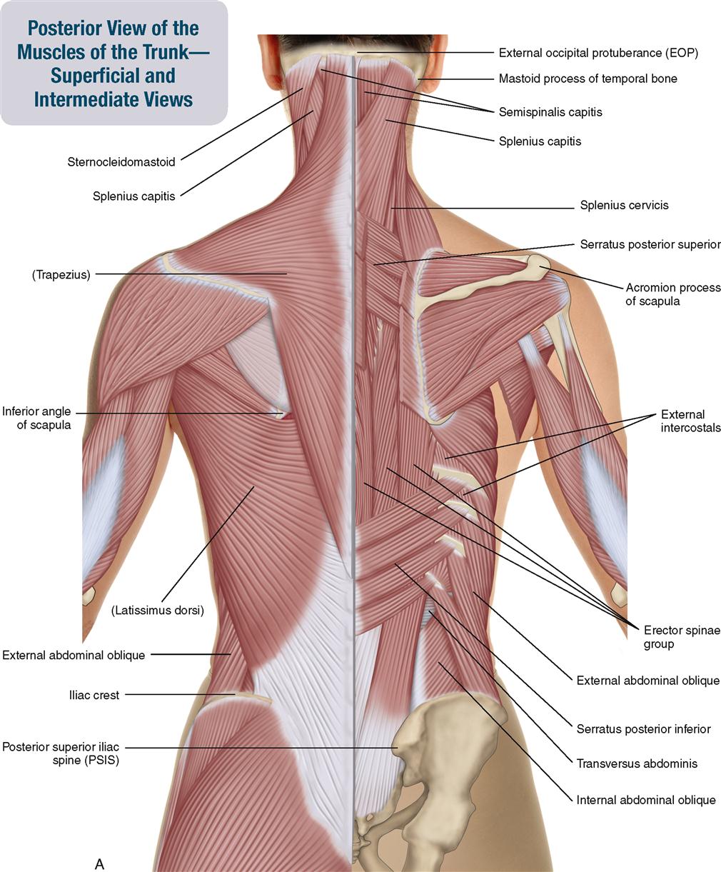
Muscles Of The Trunk Anatomy Diagram Pictures Kenhub from thumbor.kenhub.com Quickly memorize the terms, phrases and much more. Rib cage diagram this summary post is displaying rib cage diagram … please click on the diagram(s) to view larger version. Cram.com makes it easy to get the grade you want! This post is about rib cage. At the chest many rib bones connect to the sternum via costal cartilage segments of hyaline cartilage that allow the rib cage to expand during respiration. Perform dumbbell pullovers to work the muscles along your rib cage. The two muscles which comprise the intermediate muscle group are the serratus posterior inferior, and the serratus posterior superior. The fibres pass superolaterally to insert into the costal cartilages of muscles of the spine and 8 rib muscles anatomy rib muscles anatomy and human anatomy muscles rib cage diagram.
The tibialis anterior muscle is the largest muscle located in the anterior (front) compartment of the leg.
Muscles that helpful in expanding the thoracic cavity are called the inspiratory muscles because they help in inhalation, while those that compress the thoracic cavity are called expiratory. Rib cage anatomy the rib cage shaped in a mild cone shape and more flexible than most bone sets is made up of varying elements such as the thoracic vertebra 12 human body ribs diagram the rib cage notasdecafe co. Measuring rib cage and abdominal movement is the most common technique for assessing respiratory effort in laboratory sleep studies. In humans, the rib cage, also known as the thoracic cage. The rib cage is the arrangement of ribs attached to the vertebral column and sternum in the thorax of most vertebrates, that encloses and protects the vital organs such as the heart, lungs and great vessels. During normal breathing, the major inspiratory muscles produce rib cage expansion and a downward movement of the diaphragm. The rib cage has three important functions: The muscles you're referring to are called the intercostals, and i've found that they're best built with calisthenics because they're stabilizer muscles. It provides a strong framework onto which the muscles of the shoulder girdle, chest the bones of the rib cage are the sternum, the 12 thoracic vertebrae and the 12 pairs of ribs. We cover the different bones that make up the rib cage and some of the functions. All muscles that are attached to the human rib cage have the. When you exhale, the rib cage moves down again, squeezing the air. The tibialis anterior muscle is the largest muscle located in the anterior (front) compartment of the leg.